Can Stress Cause a Stroke? Yes, and Here’s What You Can Do

Can Stress Cause a Stroke? Yes, and Here’s What You Can Do

can stress cause a stroke

It is natural to feel stressed. And it might seem like there’s always something more to worry about. Stress has a function, but when it lasts for an extended period of time (chronic stress), it may do more damage than good. It may also lead to significant health problems such as high blood pressure, heart disease, and strokes.


A stroke occurs when a region of the brain does not get enough blood and nutrients. Depending on which section of the brain is affected, a stroke may induce a variety of symptoms. A portion of the face or body may feel weak. A stroke may impair speech or eyesight.


You’re going to discover how can stress cause a stroke, as well as what measures you can take to minimize stress and enhance your general health.

How can stress cause a stroke?

What occurs when you are stressed relies on the other variables you are dealing with. Stress may induce medical difficulties or operate in conjunction with other medical conditions to cause a stroke.


There is often underlying damage to the circulatory system prior to a stroke, such as weakened or restricted arteries. This is where the stroke-stress relationship emerges: persistent stress may add to artery damage and raise the risk of stroke. But how precisely does it happen?


When the body is stressed, it releases stress chemicals such as cortisol. These hormones assist the body in retaining water, salt, and sugar, therefore maintaining blood pressure and ensuring the body has adequate fuel to operate.


While this is useful for dealing with stress, it may have detrimental health consequences if the body continues in this condition for an extended length of time.


A catastrophic health incident, such as a heart attack, stroke, or being diagnosed with a disease, may also be stressful. And that stress might slow down the rehabilitation process or possibly cause new health concerns.

What research reveals about stress​

can stress cause a stroke

Increasing levels of psychological strain (such as those caused by job demands) are linked to an increased risk of both fatal and nonfatal stroke in adults.


Here are some data that address your concern about how can stress cause a stroke if it is not managed:

Blood pressure is high.

Chronic stress may weaken the arteries when it causes high blood pressure. According to 2015 research, 77% of persons who suffered their first stroke had high blood pressure.

Reactivity of blood pressure

Blood pressure reactivity occurs when blood pressure rises in response to stress. According to 2001 research, males with excessive blood pressure reactivity were 72% more likely to have a stroke. Excessive blood pressure reactivity has also been associated with atherosclerosis and hypertension, both of which are risk factors for stroke.

Habits that cause stress

According to 2012 research, people with stressful habits such as “type A personality characteristics” and “heavy use of energy-providing beverages” are at an elevated risk of stroke.

Workplace stress

Researchers in China combined data from six investigations, including 138,782 individuals, for one study. The researchers investigated the relationship between occupational stress and future stroke risk. The research, published in the journal Neurology, demonstrates that stress-related stroke is a real possibility — and that any stress is damaging to your health and should be addressed carefully.


A 2010 research found ten risk variables that are linked to 90% of the risk of specific forms of stroke. Many of these may be triggered by work-related stress, including depression, hypertension, bad eating habits, smoking, insufficient time for exercise, or alcohol addiction.

What are the symptoms of chronic stress?

can stress cause a stroke

Chronic stress has an impact on both the mind and the body. It causes both physical and psychological symptoms, which might impair a person’s ability to function properly in everyday life.


The intensity of these symptoms varies from person to person. The following are some of the most prevalent symptoms of chronic stress:

  • Pains and aches
  • Reduced energy
  • Sleeping problems
  • Thinking that is disorganized.
  • Fatigue
  • Feelings of helplessness
  • Helplessness feelings
  • Infections and diseases are common.
  • gastrointestinal issues
  • Headaches
  • Irritability
  • Muscle tenseness
  • Anxiety and nervousness
  • Concentration issues
  • Stomach ache

If you recognize any of these scenarios or symptoms, you may be suffering from chronic stress. 


So now, we will then look at techniques to assist manage it.

Methods for dealing with chronic stress

Because there is a significant link between stress and stroke, it is critical to identify measures to minimize stress and enhance general health.


Here are a few evidence-based stress-reduction techniques:

Improve your stress resistance.

Long-term healthy habits may also be beneficial since they can help you develop resilience and allow you opportunities to relax on a regular basis. This might help you avoid being so anxious that you don’t know how stressed you are, preventing you from taking action to minimize stress in your life. It may also protect you from the more serious consequences of prolonged stress.

Work out multiple times each week

can stress cause a stroke

Exercise is an effective stress-reduction strategy since it provides two advantages. For starters, exercise increases endorphins (the brain’s “feel good” chemicals) and reduces the emotional impacts of stress. Second, exercise improves cardiovascular health, therefore protecting your body from the negative impacts of stress, such as high blood pressure.

Keep an eye on your caffeine use.

A cup of coffee in the morning may be beneficial to your health, but avoid consuming too much or too late. Too much caffeine, whether from coffee, energy drinks, or other sources, may induce jittery, anxious sensations that can add to the stress. Caffeine use too late in the day may also lead to sleeplessness. Pay attention to the dose and timing of your caffeine consumption if you suffer from anxiety or sleeplessness.

Recognizing the relationship between stress and stroke

Can stress cause a stroke? YES. Chronic stress has a direct influence on the cardiovascular system, causing artery damage if left untreated. A stroke is a medical emergency caused by a blocked or ruptured artery in the brain. Vascular disease may ultimately lead to a stroke.


Chronic stress is most usually caused by the job, relationships, or financial difficulties. While we cannot always control certain circumstances, we can influence how we react to them. Selecting activities like exercise and deep breathing may help relieve stress and, as a consequence, lower your risk of stroke.

SHARE THIS POST

Facebook
Twitter

The Best Speech Therapy Exercises for Stroke Patients

The Best Speech Therapy Exercises for Stroke Patients

Best Speech Therapy Exercises

Strokes, also known as cerebrovascular accidents (CVA), may impact and damage one or more brain parts. A person who has had a left-brain stroke may have difficulty communicating. Speech therapy exercises for stroke patients aid in overcoming communication difficulties.


Speech therapy exercises can aid in the recovery of speech after a stroke. You’ll learn some fantastic speech therapy exercises you can do at home. Then, even if you can’t speak, we’ll give you some pointers to get you started.
The Advantages of Speech Therapy Exercises

Speech therapy activities are excellent for treating language impairments such as aphasia. These problems develop when the brain’s language center is injured as a result of an accident such as a stroke.
 

The language center is located in the brain’s left hemisphere. As a result, aphasia often arises after a left hemisphere stroke or brain damage.


Speech therapy activities promote neuroplasticity, the brain’s capacity to remodel itself, which helps enhance language abilities. Healthy parts of the brain may take over language function through neuroplasticity.


Speech therapy exercises train new brain parts to perform speech and communication functions.

At-Home Speech Therapy Exercises

You should ideally work with a Speech Language Pathologist to enhance your language abilities. In the interim, you may do the following speech therapy activities at home.


Here are some home speech therapy exercises to try:

Tongue Out and In

speech therapy exercises

Stick your tongue out for a few seconds, then draw it back in. Hold for 2 seconds before repeating. This helps educate your tongue to move in coordinated patterns, resulting in improved speech.

Side-to-Side Tongue

Open your mouth and move your tongue to the right corner of your mouth for this speech therapy practice. Touch the left corner of your mouth after a few seconds. Hold for 2 seconds before repeating.


Up-and-down Tongue

Open your mouth and extend your tongue. Then, bring your tongue up to your nose. Hold for a few seconds before extending your tongue toward your chin.


All of these speech therapy exercises should be done in front of a mirror so that you can gain visual feedback.

Make Your Kiss Face

After mastering the grin:

  1. Try creating kissy looks by ping your lips.
  2. Put your lips together, and then relax.
  3. Repeat as many times as you can.

Slowing down the movement will give you more control.

Repetition of Consonant and Vowel Pairing

speech therapy exercises

Now that we’ve accomplished the basic speech therapy exercises let’s move on to more sophisticated tasks.

Choose a consonant you have difficulty pronouncing and couple it with each vowel.

Sentence Creation

Patients suffering from speech apraxia have no difficulty with the cognitive aspects of language production. Their capacity to move their lips and tongue, however, is hindered.


As a result, reading aloud allows you to practice speaking. Begin by practising a phrase or two for little periods, such as one or two minutes. Then, gradually increase your practice time.

Phonological Transformation

Phonology-related speech therapy activities may assist patients in enhancing their capacity to create speech.


You will estimate the number of syllables in a word for this activity. Request that a caregiver sits with you and speak various words. Guess how many syllables a word has each time they speak it. This practice is therapeutic in part because of the feedback.

Word Puzzles

Word games are excellent adult speech therapy exercises. Even though you are not making a speech, these games test your language processing abilities.


Try word games like word searches or crossword puzzles to improve your problem-solving and visual processing skills.


When you play most brain games regularly, they will help you enhance your speech.

speech therapy exercises

What Should You Do If You Can't Talk At All?

Patients unable to speak after a stroke have most certainly suffered significant damage to the brain’s language area. Recovery will need more time and effort, but it is often doable.


Most speech therapy activities are inaccessible to mute patients, but they may take a step back and attempt a new speech treatment: singing therapy.


Surprisingly, even if a person cannot speak, they can generally sing their words. This is because speaking is a left-brain function, while singing is a right-brain one.


Singing therapy, a novel technique, assists patients in capitalizing on this potential for rehabilitation.


Many persons who struggle to regain their speaking ability after a brain injury or stroke find remarkable progress with singing therapy.

Should You Seek the Help of a Speech-Language Pathologist?

Doing these workouts alone every day might get tedious. This occurs when you do not get any progress reports or feedback. You may end up performing them incorrectly.


If you or someone you care about has had a stroke and has moderate to severe communication issues, you may choose online speech therapy.


If you have difficulty with your speech and cognitive abilities, you should consider working with a speech-language pathologist (SLP).


An SLP is qualified to assist you in regaining your speech after a neurological injury, and they deal with all of the aspects of speech recovery that we discussed before.


Most individuals work with an SLP for as long as their insurance allows, then switch to speech apps when their insurance runs out.

speech therapy exercises

Which problem areas do you need attention for?

Speech functions that your SLP will evaluate include speaking, listening, writing, reading, and remembering, among others.


Because oral motor skills are just a minor portion of the total speech, working with a professional and using speech therapy apps might be beneficial.


Your SLP will examine your problem areas and create a treatment program tailored to your specific requirements.

Performing the Most Effective Speech Therapy Exercises

Speech and communication do not begin and end with the exchange of words. It entails knowing what to say, how to say it, when to breathe, and how to make whole sentences.


Speech therapy activities may be quite beneficial to stroke sufferers. These may assist children in learning how to create sounds, form words, and appropriately breathe during speaking.


Speech difficulties may be quite irritating, particularly when they do not appear to improve with time. Working with a professional speech pathologist is the greatest approach to achieving visible, long-term speech rehabilitation.

SHARE THIS POST

Facebook
Twitter

Hand Therapy Ball Exercises to Improve Fine Motor Skills

Hand Therapy Ball Exercises to Improve Fine Motor Skills

hand therapy ball exercises

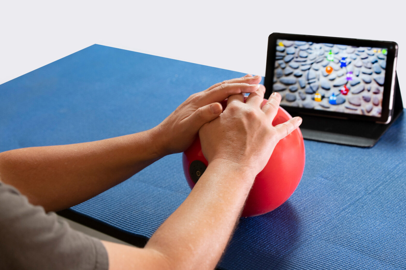
Hand therapy ball exercises are a fantastic and cost-effective choice for those who have had a stroke and want to regain strength and agility in their hands.


Because they are not complicated and can be used in various ways, hand exercise balls make it easy to do a diverse array of useful workouts designed to strengthen the hands.


This article will discuss hand therapy ball exercises that have proven to be the most successful in enhancing fine motor skills after a stroke.

Effective Hand Therapy Ball Exercises

Practices that include a lot of repetition are the most helpful when someone has had a stroke to speed up healing in the hands. The brain will be stimulated, and its capacity to rewire itself will be promoted if one consistently practices these effective hand therapy ball exercises.


You should begin with one set of 10 repetitions of each exercise; then, as your strength improves, you should aim to do more sets. You should try to perform these exercises multiple times per day if you are not experiencing any pain from doing them.

Power Grip

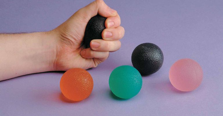
This hand therapy ball exercise can help improve your grip so that it is simpler to hold items, lift them, and release them. Stimulating the ball will focus the flexor muscles to build strength for picking objects up. It will also be crucial to practice dropping the ball, stimulating the extensor muscles that enable you to let stuff go from your hand. In relieving joint discomfort, this activity also helps alleviate tension and anxiety.


Put your fingers and thumb around the therapy ball and squeeze it as if you were making a fist. After applying pressure for a moment, let go of the ball and spread your fingers as far as possible.

Pinch

hand therapy ball exercises

You should grasp the hand therapy ball using your extended fingers and thumb for this particular exercise. And by extended, we mean maintain all your fingers straight. This will assist in strengthening muscles different from those targeted when the fingers are curled.


Pinch the ball for a longer time to make the hand therapy ball exercise that you are doing more difficult.

Flexion and extension of the thumb

The thumb is crucial in different hand tasks like pinching and gripping. Thus it’s necessary to develop its muscles to control better.


Place the therapy ball on your palm while keeping it as flat as possible, using your thumb to hold it in place. Your palm should be in a flat position.


After that, roll the ball up and down your palm using your thumb to move it. This motion focuses almost exclusively on your thumb.

Roll on table

Put your hand on top of the hand treatment ball on a table, and then put the ball down.


The next step is to roll the ball from the bottom of your palm to your fingers while maintaining a flat hand.


If you apply an insufficient amount or an excessive amount of pressure, the ball will be difficult to manipulate. Because of this, individuals can practice adjusting and maintaining a certain amount of pressure on the ball when participating in this hand therapy ball exercise.

Finger Flexion

hand therapy ball exercises

Unlike the Power Grip exercise, you won’t be utilizing your thumb in this hand treatment ball exercise.


Instead, hold the therapy ball in your hand and push onto it with all your fingers, save your thumb in the interest of the press.


Take note of how much more difficult it is to squeeze the ball when you cannot use your thumb. The muscles that enable you to flex your fingers will get stronger due to this exercise.

Thumb Roll

During this hand treatment exercise, your thumb will be targeted specifically, and you will be encouraged to move it through its full range of motion. As a consequence, it will help avoid stiffness and increase control.


Put the treatment ball on the palm of your hand. Maintain as much flat a surface as possible with your palm, and use your thumb to keep it in place. After that, roll the ball around in a circle on your palm by using your thumb to guide it.

Finger Squeeze

Individuals can strengthen the muscles that adduct their fingers by working with the hand treatment ball. These muscles enable you to draw the fingers together, which are significant in gripping items.


Put the treatment ball between your two fingers and gently squeeze them together to compress it. Then let go of the pressure. You may perform this between various combinations of fingers, so be sure to work out all your fingers!


It is important not to forget about any of your fingers, especially the ones that are more difficult to work with, such as your ring and pinky fingers.

Thumb opposition

hand therapy ball exercises

This exercise uses a therapeutic ball and is very similar to the Thumb Roll; however, instead of rolling the ball in a circular motion, you will be rolling it from side to side.


Start by putting the treatment ball on your palm and use your thumb to hold it in place. The next step is moving the ball from left to right using your thumb.

Hand Therapy Ball Exercises Offer Numerous Benefits

It is essential to modify the hand treatment exercises to meet the objectives you have set for your rehabilitation.


Some individuals may need these exercises to strengthen their hands, particularly those individuals who like engaging in handicrafts.


When there is a possibility of neurological damage, the goal of striving to enhance resistance should not be the only focus of treatment. Instead, the emphasis should be placed on repetition, which gives the brain the stimulus it requires to reorganize itself and make new neural connections.

Hand exercises can improve the quality of life.

The capacity to use both hands to grip and let go of items, type on a computer, button a blouse, or even write a message to someone you care about is crucial to having a good quality of life. If your stroke has robbed you of this ability, take action to improve your quality of life by beginning an at-home exercise program. As you work to retrain your brain following the neurological damage caused by your stroke, these finger and hand exercises for stroke recovery can assist you in regaining the use and skill of your hands.

SHARE THIS POST

Facebook
Twitter

Cerebellum Brain Damage: Causes, Effects, and Rehabilitation

Cerebellum Brain Damage: Causes, Effects, and Rehabilitation

Damage to the cerebellum causes

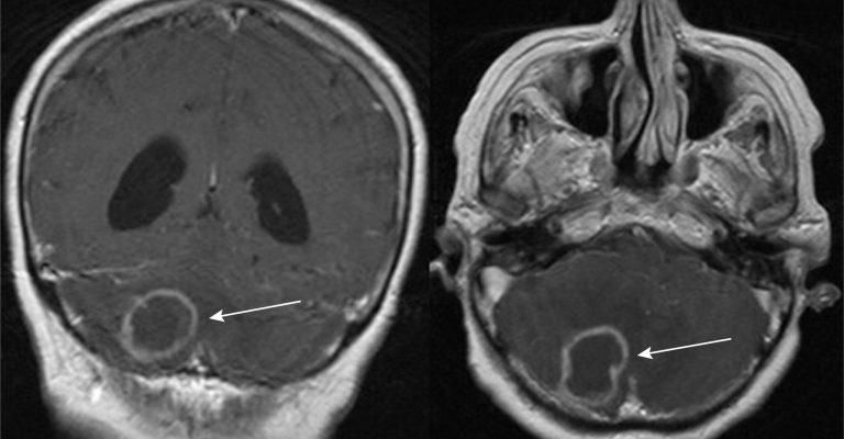
Although cerebellar brain injury is uncommon, the consequences may be severe. Damage to the cerebellum causes substantial motor, visual, and cognitive impairments. Individuals may be able to regain impaired functions with the right therapy.


In this post, we’ll look at the implications of cerebellar brain injury and therapy options to help you recover.

What exactly is the cerebellum?

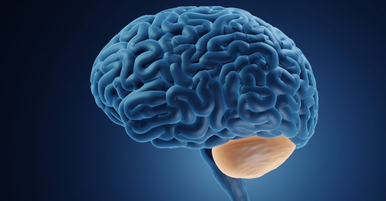
The cerebellum (Latin for “small brain”) is found at the brain’s base, just behind the brain stem. While the cerebellum only accounts for roughly 10% of the brain’s mass, it includes anywhere from 50% to 80% of the brain’s total number of neurons (brain cells).


The cerebellum coordinates voluntary motor action, balance and equilibrium, and muscle tone.


The cerebellum is responsible for movement coordination, balance and equilibrium, and muscle tone. However, it also has a wide range of cognitive, linguistic, and emotional functions.

Cerebellum brain damage causes

Anoxic brain injury, neurodegenerative illnesses, or infection are all common causes of cerebellum brain damage. Alcoholism may also wreak havoc on the cerebellum.


When the cerebellum is damaged, the signals it transmits throughout the body weaken or stop completely. This disturbance is what causes the different secondary symptoms of cerebellar injury.

Cerebellum brain damage's secondary effects

A cerebellar injury may result in a variety of symptoms. The following are some of the most prevalent secondary outcomes of cerebellar brain damage:

Damage to the cerebellum causes

Apraxia

The cerebellum transmits inhibitory or excitatory impulses to different muscle groups to fine-tune movements. Damage to the cerebellum causes the timing of instructions delivered from the cerebellum to the afflicted muscles. This causes apraxia or a loss of coordination.


Apraxia may impact any movement that needs coordination, making many ordinary tasks difficult. Many people with apraxia walk with a broad, faltering stride and take longer to accomplish basic actions.


Apraxia may also impact your face muscles and even your tongue, resulting in slurred speech and swallowing difficulties.

Motor function changes

Other motor symptoms caused by cerebellar brain loss include:


  • Dysmetria is the over- or under-estimation of the strength, distance, or speed required for undertakings.
  • Dysdiadochokinesia is the inability to produce quickly alternating motions.
  • Shaking when making deliberate motions is referred to as intention tremors.
  • Delayed movement initiation: moving at a slower rate than expected.
  • Hypotonia refers to poor muscular tone.


The extensive range of motor problems that might arise as a result of cerebellar brain loss demonstrates the cerebellum’s role in many complex motor activities.

Problems with balance

Damage to the cerebellum causes

Cerebellar injury may cause severe balance problems since equilibrium is dependent on the continued cooperation of multiple muscle groups. This might make it difficult to move, climb stairs, or shift positions.


You may have difficulty sitting or standing up straight, or you may have dizziness.

Eye movement issues

The cerebellum is not only important for coordinating and refining muscular movements in the body, but it is also in charge of managing eye movements. The eye may exhibit fast, uncontrollable movements after cerebellar brain injury, a phenomenon known as nystagmus.


After cerebellar damage, a person with nystagmus may experience three forms of eye movement:


  • Horizontal nystagmus is characterized by side-to-side eye movements.
  • Vertical nystagmus is characterized by up and down motions.
  • Rotary nystagmus is characterized by circular eye movements.


It is worth noting that the cerebellum also aids in the regulation of other visual processes, such as the vestibule-ocular reflex (VOR). The VOR is what enables you to keep viewing a steady image even as you move about.

Speech changes

The cerebellum coordinates the muscles involved in voice articulation as well as certain cognitive components of language.


Ataxic dysarthria is a frequent speech impairment related to cerebellar brain injury.


Ataxia is defined as a loss of motor control, and dysarthria is defined as difficulty coordinating the muscles involved in speech. This may result in sluggish, slurred, and/or disorganized scanning speech.

Cognitive effects

Damage to the cerebellum causes

Although cerebellar brain injury was previously assumed to solely influence motor activities, research has demonstrated that it may also disrupt cognitive processes.


You may enhance your cognitive skills by combining speech therapy activities with cognitive rehab exercises.

Treating the effects of cerebellar damage

Damage to the cerebellum causes the messages that the cerebellum is in charge of sending. Because the cerebellum is so important in motor coordination, this usually signifies that muscles aren’t getting the messages they need to coordinate movements.


Individuals must thus strengthen communication between their brain and the rest of their body in order to regain impaired functions. This is accomplished by doing repetitive workouts that promote neuroplasticity, the brain’s inherent healing process.


When you practice a skill, your brain builds neural connections linked with that activity, even if you don’t do it flawlessly. These routes will get stronger as you practice. The brain may recover its capacity to readily employ these new neural connections over time, enabling you to coordinate movement once again.

Therapy options for cerebellum brain injury recovery

Because cerebellar brain loss affects each individual differently, the therapy necessary for recovery may differ. However, most people who have suffered cerebellar brain injury benefit from physical, occupational, and/or speech treatment. Each of these treatments aims to enhance healing by activating neuroplasticity.

Physical therapy

Damage to the cerebellum causes

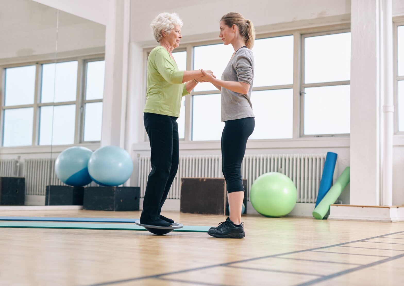
Physical therapists may concentrate on retraining the brain to improve balance and coordination. To improve functional recovery, they may employ strengthening exercises and gradually increasingly difficult tasks. Some physical therapists specialize in vestibular treatment, which may assist people with dizziness and balance issues.

Occupational therapy

Occupational therapists assist people in carrying out their regular duties. They may advise people with apraxia to practice each phase of activity independently before combining them all together. They could also suggest compensating measures and adapted equipment. Some occupational therapists specialize in vision therapy, which may assist with nystagmus and other visual issues.

Speech therapy

Speech therapists may help patients with ataxic dysarthria improve the coordination of their mouth musculature. They may also aid in the recovery or replacement of lost cognitive skills.

The more you use afflicted functions; the more your brain will establish new neural connections to encourage improvement.

Cerebellum understanding brain injury

Damage to the cerebellum causes significant issues with muscular coordination. Fortunately, there is hope for healing.


Engaging your brain’s neuroplasticity is the key to mending any brain damage, including cerebellar lesions. Daily therapeutic exercises may help you improve your balance, coordination, and cognitive abilities. Depending on the severity of your injuries, you may even heal completely.

SHARE THIS POST

Facebook
Twitter

15 Effective Exercise for Cerebral Palsy

15 Effective Exercise for Cerebral Palsy

Exercise for Cerebral Palsy

When it comes to helping someone with cerebral palsy, exercise physiology is often the first step. Exercise is helpful to everyone, but it is especially useful to persons with cerebral palsy.


Exercise for cerebral palsy can help individuals increase their degree of independence and quality of life in a variety of ways, from maintaining the body flexible and strong to enhancing mobility and avoiding injury.


Cerebral palsy patients might have either high or low muscle tone or a combination of the two. Muscle tone may also alter throughout the day for certain persons with cerebral palsy. High muscle tone may result in spasticity and rigidity, while low muscle tone results in weakness and excessive flexibility.


Different exercises for cerebral palsy are often used to increase mobility and prevent painful muscular tightness, which occasionally necessitates surgery.


We’ve listed 15 effective exercises for cerebral palsy adults below.

Joint rotations

Rotate all of your primary joints, including the neck, to increase or maintain your range of motion. Rotate them a few times in one direction, then switch to the opposing way. This will help to reduce stiffness and increase circulation.

Stretching calves

Many people with cerebral palsy have stiffness in their calves. This may cause the heels to lift, resulting in aberrant gait patterns such as toe-walking. Stand facing a wall and take a large stride back with one leg to extend tight calves. Adjust your legs such that the front leg is bent and the rear leg’s heel is lifted.


Lean your arms on the wall for support and slowly descend your heel. Hold for 20-30 seconds before switching legs.

Exercise for Cerebral Palsy

Stretching quadriceps

The quadriceps are the muscles on the front of your thighs. When your knees get stiff, they may begin to bend inwards, contributing to aberrant walking patterns. Sit on the floor with one leg bent back and the other out in front of you to stretch your quads. Lean back gently until you feel tension but not discomfort. Hold for 20-30 seconds before switching legs.

Stretching the hamstrings

The hamstrings are the muscles at the rear of your thighs. Because these muscles oppose the quads, they must also be stretched to allow for normal hip and knee motions. Sit on the floor with both legs straight in front of you and bend forward in a pain-free range to stretch the hamstrings. Hold for 30 seconds before repeating.

Shoulder flexion

Raise one arm behind your head and bend it. Gently push the elbow back with your other arm’s hand until you feel the strain. Hold for 20-30 seconds before switching arms.

Arm across the chest

Swing one arm over your chest, holding it in place with the other. Gently squeeze the arm towards your body to tighten the wrap around the chest. Hold for 20-30 seconds before switching sides.

Trunk tilts

Lay on your back and twist your upper body to one side slowly. Legs should not be moved. After 20 seconds, rotate your upper body to the opposing side. This stretch stretches the sides of your trunk.

Exercise for Cerebral Palsy

Trunk twists

Trunk twists may be done while sitting and standing. Keep your lower body steady by placing your feet on the floor. Gently rotate your upper body to one side, hold for 20 seconds, and then repeat.

Wheelchair aerobics

Wheelchair aerobics may be an efficient cardio exercise if your cerebral palsy mostly affects your legs or one side of your body. It stresses upper body mobility while sitting.

Dancing

Dancing is a workout that everyone may undertake at any time and in any place.


Turn on some upbeat music for cardio. Music has been shown to affect physical changes in heart rate, blood pressure, muscular tension, and respiration.

Wheelchair pushing

Pushing your wheelchair is a terrific arm and chest strengthening exercise, as well as a cardio workout for those with CP in the lower body.


As much as possible, push your wheelchair instead of letting others push it for you. This is a simple method for incorporating strength training into your regular routine.

Wheelchair push-ups

First, make sure your wheelchair brakes are engaged. Then, with your feet level on the floor, sit on the edge of your seat.


Lift your body up by placing your hands on the armrests and pressing down. Your feet should stay on the floor, supporting part of your weight.

Exercise for Cerebral Palsy

Resistance bands

Resistance bands come in a number of resistance levels and may be used for a variety of muscle training workouts.

Using weighted cuffs

Putting on some weighted cuffs around your wrists or ankles and carrying them about with you as you go about your day is a simple approach to increase your strength. It may not seem like much at first, but even a few more pounds can make a significant difference over time.

Aquatic workouts

Have you ever taken the time to consider how much more challenging it is to move swiftly when you are submerged in water as opposed to when you are on land?


This is due to the fact that liquids possess a viscosity (level of resistance) that is higher than that of gases. The increased resistance will assist in making the muscles stronger.

Few things to keep in mind with regards to exercises for cerebral palsy

You must realize that there is no such thing as a one-size-fits-all exercise program for those with impairments. Every individual is distinct. Whether they have a disability or not, their fitness requirements will be heavily influenced by their abilities.


Nonetheless, encouraging individuals to be active from a young age has long-term advantages in terms of helping them live healthier and more active life.

Takeaway

Exercises for cerebral palsy may be done at home with the help of loved ones, but it is still essential to have the assistance of trained specialists while you are doing your exercises.


Certain types of cerebral palsy need specialized care and attention. If your loved one has a disability, the ideal sort of individuals to help them attain the degree of fitness that you would desire for them are qualified fitness specialists who specialize in working with adults.

SHARE THIS POST

Facebook
Twitter

Elite Athlete Products, Inc. is Awarded a Phase I STTR Grant From NIH-NICHD to Advance their Rehabilitation System for Post-Stroke Gait Training

Elite Athlete Products, Inc. is Awarded a Phase I STTR Grant From NIH-NICHD to Advance their Rehabilitation System for Post-Stroke Gait Training

Elite Athlete Products, Inc. (EAP) and their academic partners at the University of Michigan and Wayne State University have been awarded a $344,843 Phase I grant from the STTR Program under the Department of Health and Human Services, National Institutes of Health: Eunice Kennedy Shriver National Institute of Child Health & Human Development (NICHD) to support a study of a post-stroke gait mobility/rehabilitation device.


Despite significant advancements in post-stroke medical treatments and rehabilitation interventions, more than half of stroke survivors cannot walk independently in the community. Current rehabilitation devices aimed at restoring walking function are either large and unaffordable or ineffective in improving walking function, creating a significant unmet need for a portable, affordable, and effective gait mobility/rehabilitation device.


EAP is working in collaboration with researchers at the University of Michigan and Wayne State University to further the development of the NewGait rehab system.


The STTR grant award expands the work performed at Dr. Chandramouli Krishnan’s Neuromuscular and Rehabilitation Robotics Laboratory (NeuRRo Lab) housed within the Department of Physical Medicine & Rehabilitation at the University of Michigan. The NeuRRo Lab aims to develop effective and efficient rehabilitation methods for individuals with neurological and orthopedic disorders.


“A key issue in stroke rehabilitation is the lack of availability of low-cost, lightweight devices that can be easily taken home to improve therapeutic dosage—an important component for recovery after stroke,” said project lead Dr. Chandramouli Krishnan, Associate Professor of Physical Medicine & Rehabilitation, Michigan Robotics, Biomedical Engineering, and School of Kinesiology. Dr. Krishnan is also a faculty in the U-M Robotics Institute, Michigan Neuroscience Institute, and the University of Michigan-Flint Physical Therapy Department.


Most commercial gait rehabilitation systems often require uninterrupted power sources, which is a significant barrier for rural communities in developing nations. Thus, there is a critical need for non-powered technologies for rehabilitation.


“The NewGait® system is a natural fit for this need, as it is a non-powered, wearable gait rehabilitation system. Hence, we are interested in exploring this as a method to restore gait function in individuals with stroke,” Dr. Krishnan said.


However, the current NewGait® device is not tailored to stroke-specific needs, and the clinical utility has not been verified experimentally. The Phase-I STTR grant will allow EAP to address these gaps by developing a stroke-specific low-cost gait rehabilitation system based on end-user feedback and musculoskeletal modeling.


“Our team is uniquely qualified to perform this research, as we have experts in biomedical and mechanical engineering, stroke rehabilitation, biomechanics and modeling, clinical trials research, and human-centered design. We also are working with a world-class rehabilitation hospital,” said Mr. Adeeko, Founder & CEO of EAP.


The project team at Michigan Medicine includes Drs. Chandramouli Krishnan (Physical Therapist, Biomechanist, Neuroscientist, and Roboticist), Edward S. Claflin (Physiatrist and Stroke Physician), Claire Kalpakjian (Biopsychosocial Scientist and Clinical and Rehabilitation Psychologist), and Alyssa Portelli (Clinical Physical Therapist). The team also includes Dr. Edward P. Washabaugh (Biomedical Engineer, Biomechanist, and Roboticist), an Assistant Professor in Biomedical Engineering at Wayne State University.


Dr. Krishnan hopes that the project will result in a low-cost system for post-stroke gait rehabilitation and will lay the foundation for developing an evidence-based rehabilitation system that could positively affect the lives of millions of stroke survivors across the globe.


Mr. Adeeko said, “We are thrilled at the opportunity to further our research efforts on our NewGait line of devices. Funding from this award will allow us to continue development and gather pilot data for a larger clinical study.”


The project is funded by NIH NICHD grant R41-HD111289, “NewGait: A Low-Cost Rehabilitation System to Improve Post-Stroke Gait.” The mission of NICHD is “to lead research and training to understand human development, improve reproductive health, enhance the lives of children and adolescents, and optimize abilities for all.”

SHARE THIS POST

Facebook
Twitter

Neurostorming: Causes, Signs, Risks, and Treatment

Neurostorming: Causes, Signs, Risks, and Treatment

Neurostorming

Individuals who have suffered a severe traumatic brain injury may go through a period of neurostorming.


Also called paroxysmal sympathetic hyperactivity, neurostorming is an acute and extreme stress reaction caused by brain injury. For instance, a person’s heart rate, blood pressure, or temperature may suddenly increase.


Comatose patients often experience neurostorming after sustaining a severe brain injury. People with brain injuries can overcome storming, regain consciousness, and fully recover with the help of medical professionals and time.


This article will address the causes, symptoms and treatment options you can explore.

What is Neurostorming?

If you’ve suffered head trauma, you may have neurostorming, a state characterized by an overactive sympathetic nervous system (SNS). The sympathetic nervous system (SNS) triggers the “fight or flight” reaction.


When triggered, the body goes into a state of heightened attention, raising the rate at which it performs essential processes like heart rate, blood pressure, and temperature regulation.


The ability to either “fight” or “flee” from a potentially harmful or stressful circumstance is what the “fight-or-flight” reaction is all about. When danger is no longer present, the body’s parasympathetic nervous system (PNS) takes over and causes a state of deep relaxation.


It does this by bringing the heart and respiratory rates back to normal.

However, the sympathetic response may become dysfunctional after a serious brain injury. The brain has been compromised. Therefore, it cannot assess whether or not the body is in danger correctly.


As a result, the sympathetic nervous system becomes more active, leading to the unwarranted release of adrenaline and other hormones into the bloodstream.


The stress reaction is beneficial in the short term, but too much of it can harm your health in the long run (which will be discussed later in this article).


Knowing the symptoms of a neurostorming episode and getting medical help quickly can prevent more severe consequences from developing.

Subscribe to our Newsletter

Neurostorming Symptoms and Signs

After sustaining a brain injury, the signs and symptoms of neurostorming might look very different depending on the individual. Neurostorming can last anywhere from a few hours to several months after an injury.


When someone has to storm due to a brain injury, they are typically in a coma and unaware of what is occurring to them. Therefore, it is helpful for loved ones and healthcare providers to be on the lookout for symptoms of neurostorming.


Symptoms of neurostorming are listed below;

  • Temperature more than 101 degrees (38.5 degrees Celsius).
  • Having an abnormally high blood pressure (hypertension)
  • If your heart rate is over 130 beats per minute, get medical attention immediately (tachycardia)
  • Tachypnea is characterized by a rapid breathing rate of more than 40 breaths per minute.
  • Sweating excessively (diaphoresis)
  • Stiff muscles in the arms and legs
  • Back-arched neck and spine and pointing toes (abnormal posturing)
  • Restlessness
  • Increased pupil size
  • Glycogenolysis
  • A higher resting metabolic rate

Causes of Neurostorming

Neurostorming
Neurostorming

Hydrocephalus, intracerebral haemorrhages, and hypoxic brain damage are all common outcomes of a storm. However, those who have suffered traumatic brain injuries are more likely to experience this. The neurological phenomenon known as “neurostorming” may occur in 15-33% of comatose individuals with severe traumatic brain injuries.


After a brain injury, most people’s storming episodes happen by themselves and without provocation. While there are many potential causes, some of the more common ones are:

  • Modifications to pharmaceutical regimens or the abrupt discontinuation of treatment
  • Repositioning
  • Stimulation from the environment (such as alarms or other loud noises)

Repercussions of Neurostorming

The effects of neurostorming can worsen a person’s health if they aren’t treated. The dangers of untreated neurostorming include:

  • Dehydration
  • Degeneration of muscle tissue
  • Weight loss
  • Pressure ulcers
  • Injury to the skeletal muscle
 

As was previously noted, neurostorming can cause considerable increases in one’s metabolic rate and core temperature, blood pressure, and muscle tone.


A person’s energy needs can increase by as much as 100% during a storming event. Muscle mass is lost along with fat when the metabolism is increased so dramatically.


It is more likely that pressure sores will form on people whose metabolisms are speeding up and who also perspire heavily. This can be extremely painful, put you at greater risk for infection, and severely restrict your range of motion.


Those with aberrant posturing for an extended period without treatment may also suffer irreversible musculoskeletal injury.


As a result, prompt and efficient treatment of neurostorming following brain injury is essential.

Treatment

Neurostorming

Drugs are typically used to treat secondary consequences of neurostorming, such as elevated blood pressure and fever. These pills are designed to calm the body and lessen the effects of stress. Potentially avoidable secondary effects of storming may be avoided if the indications and symptoms of a hyperactive sympathetic nervous system are managed.

Neurostorming medications include, but are not limited to, the following:

  • Opioids (morphine, fentanyl, etc.)
  • Medicines administered intravenously (propofol)
  • Beta-adrenergic antagonists (propranolol, metoprolol, etc.)
  • α2-agonists (clonidine, dexmedetomidine, etc.)
  • Benzodiazepines (diazepam, lorazepam, etc.)
  • Neuromodulators (gabapentin, baclofen)

The sudden discontinuation of a drug can trigger neurostorming. For this reason, it is best to taper off medications over time as the patient improves.

Increasing caloric, protein, and water consumption may also be part of the therapy plan to compensate for the high metabolism and lessen the likelihood of muscular atrophy and dehydration.

How to Avoid Neurostorming?

Intense mental agitation, known as “neurostorming”, can occur after a head injury. When neurostorming is prevalent, loved ones may worry that their relative or friend’s health is getting worse. Storming, however, is a typical response to a traumatic brain injury of sufficient severity.


Even still, many onlookers of a neurostorm report feeling helpless. Lucky for you, there are a few ways to pitch in: Learn something and share it with others. Recognize the symptoms of neurostorming following a brain injury (such as the ones listed above).


The nurses are on high alert. If you recognize the signs of neurostorming, you can call for help before the condition worsens. You should contact a nurse if you notice changes in your loved one’s temperature or heart rate.


Make sure you’re being proactive. At last, you have options for averting neurostorming. Effective techniques include speaking softly to them, rubbing their arms and legs, and using a cool cloth to reduce their body temperature.


Your loved one’s brain can still respond to stress even if unconscious. The room should be a peaceful retreat for them, so make an effort in that direction.


How to avoid neurostorming? Examine the causes and possible symptoms of neurostorming to protect your loved ones. Explore the treatment options for neurostorming recovery.


SHARE THIS POST

Facebook
Twitter
Subscribe to our Newsletter

How to Fix Curled Toes: 6 Treatment Options That Work!

How to Fix Curled Toes: 6 Treatment Options That Work!

How to Fix Curled Toes

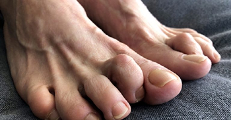
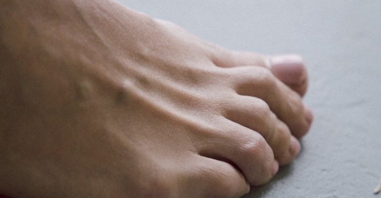
As every human being is same yet different from each other in form of shape, skin, height and other characteristics, so is our toes. Our toes are supposed to be flat and flexible against ground so they move and spread openly. They help us in walking and balancing on ground. However, curled toes are toes curling inwards or bending downwards. This condition is painful and should be treated timely before it gets worse. However, there are many causes that lead to your toe curling inwards. To treat the following condition, it is important to know the symptoms and causes for the condition as lifestyle, shoes and different things affect our feet.

Types

The types of deformed curled toes are as following.

The abnormal bend exists in the last joint of the toe which is closer to toe nail.

It has a bend that is abnormal in the middle joint toe. In this deformity, the second toe is the most affected. The reason why it’s called hammer toe is because it resembles the hammer as the toe is curling inward.

Claw Toe

The abnormal bend exists in the middle and last joint of a toe which is closest to the toe nail. It can also be due to your feet’s weakened muscles. This can affect your one or more than one toes.


There are different causes that can lead to your toes curling inwards. Root cause can be imbalance of muscles in digits. The toes become stuck initially flexibly and then rigidly, as the muscle that straightens them becomes too weak to oppose its partner. Other reasons are as follows;

Misfit shoes

Wearing shoes that are either too tight or does not fit properly leads to stress on your toe muscles that must be straight and relaxed. Tight shoes can squeeze and bend the toe. Moreover, wearing heels also put pressure on your toes which can cause this condition.

How to Fix Curled Toes

High Obesity

Severe obesity causes pressure on toes causing strain on toe muscles. A study reveals that obesity with men is cause of claw toe. 


  1. Genetics
  2. Toe injuries

A severe foot injury can cause your toe to curl inward.

Other reasons for curled toes are strokes or nerve damage, arthritis and diabetes that can cause systematic conditions and many more. However, curled toes should be treated as it can lead to complications, difficulty and pain in mobility.

Subscribe to our Newsletter

Treatment

The treatment for the condition can be as follows;

Fit Shoes

Buy shoes that are comfortable and perfect fit. Make sure the shoes are not tightly fitted so they put pressure on any toe muscle. Moreover, avoid high heels so it does not cause stress on toes and make the condition worse. It is important to wear good supportive shoes in this condition to prevent it from getting worse. Moreover, you can also use roomy shoes that will help in straightening up your toe as they will give a room for your toes to wiggle. Shoes with bigger toe space will help in such conditions.

Exercises

The better way to treat curled toes are exercising at home or in any Rehab. There are certain stretches that must be added to your routine in order to deal with your toes that are curling inwards.

Toe Lift

Toes should be raised off the ground and lower and this should be repeated 10-20 times. If you can’t move the toe in the beginning, don’t worry and take it slow and start with slight movement.  

photo-1434973539530-5538b4681aac

Stretch your toes

Try to stretch your curled toe as much straight as you can and hold for 5 seconds. If it is painful, try short repetitions and gradually increase reps.

Marble Pickups

Place few marbles on floor and try to pick them up with your toes. It is going to be challenging so you can for assistance.

Towel curl

A towel must be placed on ground and try to scrunch it with your toes. If you can’t do it properly in the first place, it’s okay, you will be better with practice and time.

Use of Splints

Use any orthopedic device that will help in keeping your curled toe in a straightened positioned with either a toe wrap, toe straighteners or any device. If you get catch on the curled toe in initial stage, you can get it straight with a tape or splint. Moreover, toe separators can help in reducing the pain.


Surgery

If any of the treatment does not help and it gets worse or permanently curled, then surgery is the only option to get your toe straight in position. The doctor can provide better details considering your condition to relieve your pain.


Curled toes can be treated by adopting a particular lifestyle in which you wear comfortable shoes and do home treatments as mentioned above. These initial care steps can save your toe from going towards a surgery procedure. However, if the toe is curled inwards permanently then surgery is the only option left.


The details regarding the surgery will be advised by the doctor according to your condition. Toes are a crucial part of our body since they help us in running, walking and mobility so if they are disturbed, it can be a problem. It also leads to painful consequences like blisters, severe pain and difficulty in mobility.


This condition should not be ignored since it can get worse rather it should be treated properly. If it is not treated timely, it can affect other toes as well and deform them. So in order to save your toe from getting to the worst condition, try to catch it up in initial stage before the inward curled toe becomes permanent and starts affecting other toes. It is better to catch it up before it’s too late.  


Learn about the various types of curled toes, and what cause them. Explore the various treatment options available.


SHARE THIS POST

Facebook
Twitter
Subscribe to our Newsletter

AFO Brace for Foot Drop: How to Find the Perfect Fit?

AFO Brace for Foot Drop: How to Find the Perfect Fit?

AFO Brace for Foot Drop

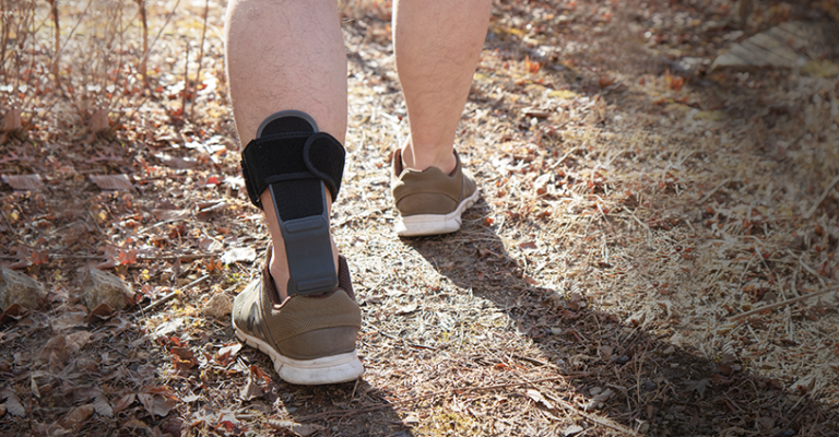
When it comes to mobility and independence, our feet play a crucial role. However, foot drop, a condition that causes weakness or paralysis of the muscles in lifting the front part of the foot, can significantly impact our ability to walk and perform daily activities. Fortunately, an Ankle-Foot Orthosis (AFO) brace offers a solution by providing support, stability, and improved gait for individuals with foot drop.


But finding the perfect fit for an AFO brace can be daunting. In this blog post, we will guide you through finding the ideal AFO brace for your needs. We’ll provide you with essential information to ensure that you find the perfect AFO brace that maximizes mobility and enhances your quality of life.


Let’s embark on this journey to discover how to regain control and confidence in your steps with the right AFO brace for foot drop.

What is Foot Drop

Foot drop is a medical condition characterized by difficulty or inability to lift the front part of the foot, leading to an abnormal gait and potential tripping hazards. It is often caused by underlying nerve or muscle damage, such as nerve compression, muscular dystrophy, stroke, multiple sclerosis, or injury to the spinal cord. Foot drop can result in dragging the foot along the ground or compensatory movements like lifting the leg to clear the foot.

AFO Brace for Foot Drop

Signs & Symptoms

Foot Drop can occur due to various underlying medical conditions or injuries affecting the leg and foot’s nerves, muscles, or bones. The signs and symptoms of foot drop may include:


Difficulty lifting the front part of the foot: The primary symptom of foot drop is an inability to dorsiflex the foot, which means raising the foot and toes upward towards the shin.


Foot dragging or slapping: When walking, a person with foot drop may have an obvious dragging or slapping gait due to the foot not being lifted high enough to clear the ground.


Tripping or stumbling: The foot may catch on the ground or objects, leading to frequent tripping or stumbling while walking.


High-stepping gait: To compensate for the dropped foot, individuals may lift the knee excessively high or swing the leg outward exaggeratedly while walking.


Weakness in the lower leg or ankle: There may be a noticeable weakness in the muscles of the lower leg and ankle, making it challenging to control foot movements.


Numbness or tingling: Some individuals may experience numbness or tingling in the foot or toes, which can accompany foot drop in some instances.


Muscle atrophy: Prolonged foot drop can lead to muscle wasting or atrophy in the lower leg, particularly in the muscles responsible for foot dorsiflexion.


Difficulty with balance and coordination: Foot drop can affect overall balance and coordination, as compensatory movements may be required to maintain stability while walking.

Why Is It Necessary to Wear an AFO Brace If You Suffer from Foot Drop?

Foot drop occurs in around 20% of people who survive a stroke. If you have this issue, your foot may “drop” or drag as you walk because you have trouble dorsiflexing. When people walk, their brains often tell their muscles to avoid letting their toes dangle. Injuries to the brain or spinal cord may interrupt the signals that typically send signals to the muscles, leading to foot drop.
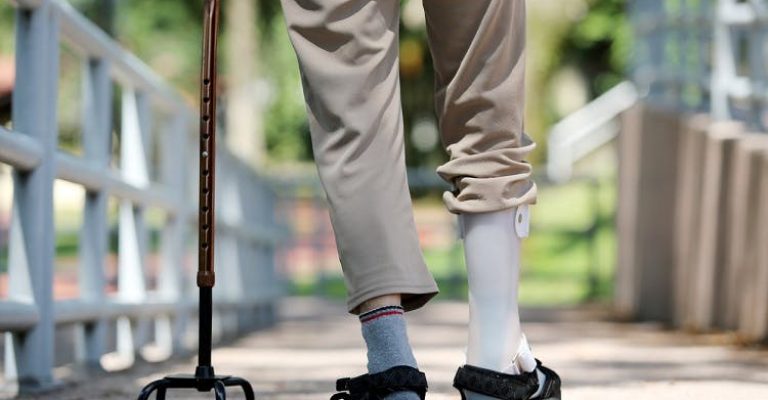
 

A significant safety issue might arise from a lack of toe-lifting capacity. If your toes are dragging, you can trip over a rug or a fissure in the pavement. In addition, numbness of the afflicted foot is a common symptom of foot drop. This makes it more likely that they might trip and tumble if they walk on rough ground.

AFO Brace for Foot Drop

Some people with foot drop prefer to keep from tripping their toes by swinging that affected limb out to the left with each stride. Some people do this by extending their legs in a walking motion to get their foot off the floor.


Changing to a new walking style is not likely to aid in rehabilitation from foot drop, and it does little to fix the underlying source of the condition. The flip side is that they may lead to even more severe side effects, such as persistent pain and increased exhaustion.


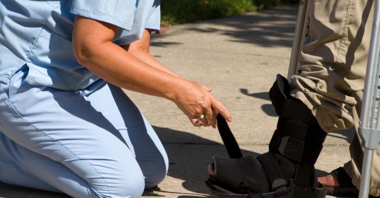
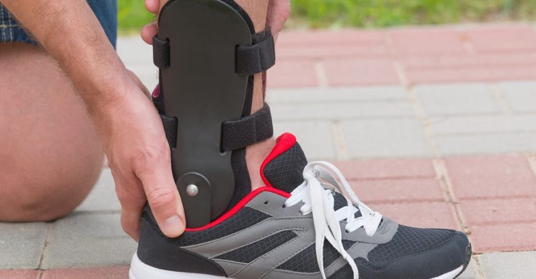
For this reason, if your child has foot drop, your doctor or therapist may recommend an AFO brace—ankle foot orthoses aid in stabilizing the foot and preventing the toes from dragging. You’ll feel more secure on your feet and won’t have to worry about developing unnatural gait patterns. This allows people with foot drops to go about more comfortably and effectively.

AFO

When choosing an ankle-foot orthosis (AFO) brace for foot drop, it’s essential to consider several factors to ensure the brace meets your specific needs. Here are some key aspects to look for:

Fit and Comfort

The brace should fit properly and be comfortable for extended periods. Look for adjustable straps or sizing options to achieve a customized fit. Padding and cushioning in critical areas can enhance comfort and prevent irritation or pressure points.

Rigidity and Support

The AFO brace should offer sufficient rigidity and support to hold the foot in a dorsiflexed position. It should effectively prevent foot drop and allow for proper clearance of the toes during the swing phase of walking. The level of rigidity required may vary depending on the severity of foot drop and individual preferences.

Material and Durability

Consider the material of the brace. It should be sturdy and durable to withstand regular use and provide long-lasting support. Common materials used in AFO braces include plastic, carbon fiber, or a combination of both. The choice of material may depend on weight, durability, and flexibility.

Range of Motion and Adjustability

Look for an AFO brace that allows for an appropriate range of motion at the ankle joint. It should facilitate natural movement while providing the necessary support. Additionally, consider whether the brace offers adjustability to accommodate swelling, muscle tone, or foot size changes over time.

Breathability and Moisture Management

Proper ventilation and moisture-wicking properties are essential to keep the foot dry and comfortable. Look for AFO braces with breathable materials or features promoting airflow and reducing skin irritation or bacterial growth risk.

Ease of Use

The AFO brace should be easy to put on and take off independently or with minimal assistance. Consider features such as fasteners, straps, or closures that are user-friendly and secure.

Compatibility with Footwear

Ensure the AFO brace is compatible with your regular footwear. It should fit comfortably inside your shoes without causing excessive pressure or discomfort. Some AFO braces are designed to be worn with specific types of shoes, so consider your footwear options when choosing a brace.

Using a foot drop brace long-term can have some drawbacks and considerations. Here are a few disadvantages:

Muscle Atrophy

Relying on a foot drop brace for an extended period may result in muscle weakness and atrophy. Since the brace provides external support to lift the foot, the muscles responsible for dorsiflexion (lifting the foot) may become underutilized and weaken over time.

Dependency

Long-term use of a foot drop brace can lead to dependence, where the individual becomes reliant on the brace for walking or performing daily activities. This can hinder the affected muscles and nerves’ natural recovery and rehabilitation process.

Decreased Sensation

Foot drop braces often provide rigid support and may limit the natural range of motion of the foot and ankle. Prolonged use of such braces can reduce sensory feedback from the foot, which is important for balance, proprioception, and detecting ground surfaces. Reduced sensation may increase the risk of falls and accidents.

Skin Irritation or Pressure Sores

Continuous use of a foot drop brace can cause skin irritation, pressure points, or even pressure sores, especially if the brace is ill-fitting or not correctly adjusted. It is essential to regularly monitor the skin’s condition and make necessary adjustments to prevent discomfort or skin damage.

Psychological Impact

Long-term use of a foot drop brace may psychologically affect the wearer. It can constantly remind them of their condition, potentially leading to frustration, self-consciousness, or decreased self-esteem.

Inconvenience

Wearing a foot drop brace constantly can be inconvenient in certain situations. It may limit the choice of footwear and require extra time and effort to put on and take off. Activities such as swimming or showering may be challenging with the brace.

An AFO brace is often a crucial defense stage when a patient presents with foot drop. While an AFO may be helpful, it is meant to be a short-term safety measure and not a long-term solution. 


As part of a comprehensive physical therapy program for your whole body, you should immediately begin doing foot drop exercises. Optimal neuroplasticity and subsequent foot and ankle recovery will result from this. Neuroplasticity allows you to reduce reliance on an AFO as your progress continues gradually.

SHARE THIS POST

Facebook
Twitter

Scissoring Gait and Cerebral Palsy: Causes, Risks, & Treatment

Scissoring Gait and Cerebral Palsy: Causes, Risks, & Treatment

Scissoring Gait and Cerebral Palsy

Have you ever heard of the term spastic cerebral palsy? Do you know what it is?


Damage to the brain before, during, or shortly after birth causes spastic cerebral palsy, a developmental condition. This disorder disrupts the growth of motor skills. Spastic CP is characterized by jerky motions, muscular tightness, and joint stiffness.


Cerebral palsy can make even the most basic activities, like walking or picking up a small object, difficult.


As a result of severe brain injury, some children with spastic CP also acquire secondary disorders.


Attention Deficit Hyperactivity Disorder (ADHD) and epilepsy are two examples of conditions that frequently occur together.

Spastic CP Subtypes

One’s specific brain injury and associated symptoms can be further specified under each of the four primary categories of cerebral palsy.


Spastic cerebral palsy is broken down into subtypes according to where in the body the patient is having trouble moving.


Muscle stiffness in one or both arms, both legs, or even one entire side of the body is common in children with spastic CP.

Diplegia Spastica

When two limbs are affected by spasticity, the legs are most often affected. Some children with diplegia may also have difficulty moving their arms and shoulders.


Diplegia is a common complication of cerebral palsy, usually resulting from premature delivery.

The complete side is affected when one side of the body is affected by spastic hemiplegia.


In most cases, the arm is injured more than the leg and can be identified by a stiffly flexed wrist or elbow.


Hemiplegia is a neurological condition caused by bleeding in the brain during pregnancy.

Quadriplegia with Spasticity

Disorder of the spinal cord causing spasticity in the limbs.


When all four limbs are affected, it’s called spastic quadriplegia. In most cases, the legs suffer more damage than the arms.


Facial muscular control may be impaired in people with quadriplegia.

Scissoring Gait and Cerebral Palsy

Aetiology and Precipitating Factors

The term “cerebral palsy” is used to describe a wide range of movement abnormalities that can be caused by damage to the developing brain. Different forms of cerebral palsy result from harm to various brain regions.


Damage to the brain’s motor cortex or the pyramidal tracts, which run from the motor cortex to the spinal cord, is the root cause of spastic cerebral palsy.


Damage to the motor cortex and pyramidal tracts leads to spasticity and understanding how these systems work might shed light on this condition.

Cortical Dysfunction in the Motor System

The cerebral cortex, the bulk of the brain, houses the motor cortex.


Several subregions of the brain’s cortex, collectively known as the motor cortex, are in charge of communicating with the brain’s other regions to regulate movement.


The motor cortex’s role in controlling overt motion is crucial to understanding the impact of cerebral palsy on a person’s daily life.


When this part of the brain is damaged, voluntary movement becomes less coordinated and fluid, leading to spasticity.

Injury to the Pyramidal Tracts

When sending and receiving messages between the brain’s cortex and the spinal cord, the brain’s pyramidal tracts serve as the highways.


Disruption of the pyramidal tracts prevents the motor cortex from sending accurate signals to the spinal cord.


The spinal cord and the brain and brain stem make up the CNS. Visual, tactile, and motor abilities all originate in these brain regions.

Symptoms and Signs

Each child with spastic cerebral palsy experiences the disorder in a unique way. The degree of the child’s brain injury and any co-occurring disorders will determine the range of symptoms they experience.


In most cases, the following are the most noticeable signs of spastic CP:


  • Muscle rigidity (hypertonia) on one or both sides of the body, accompanied by a lack of flexibility and resulting in accentuated gestures
  • Constricted movement
  • Unnatural walking pattern
  • Knees crossed
  • Inability to fully stretch joints
  • On the tips of one’s toes
  • Contractures
  • Inappropriate Reactions
  • Co-occurring disorders, such as hearing and visual impairment, may also present themselves, but these are caused by the initial birth injury rather than the cerebral palsy itself.


Cerebral palsy is not always evident in a child’s early years. Symptoms don’t usually show up until a child stops meeting conventional developmental markers.


Many children have the jerky reflexes seen in individuals with spastic CP when they are toddlers. Up to age 5, a definitive diagnosis of cerebral palsy may not be made.

Scissoring Gait and Cerebral Palsy

Spastic CP treatment is situational. Treatment plans are influenced mainly by the intensity of symptoms, the location of mobility issues, and the presence of any secondary disorders.


Physical therapy, occupational therapy, speech therapy, medication, and surgery are the five basic ways to treat CP.


The Benefits of Physical Therapy

Spastic CP is the most common form of the disorder, and physical therapy is often the first line of defence.


When it comes to physical treatment, the end goal is always maximum autonomy for the kid.


Therapeutic stretches and movement are emphasized throughout this procedure. Learn more about physical therapy here.

Professional Counseling for Occupational Performance

Children with spastic cerebral palsy may also benefit from occupational therapy. An individual’s independence in completing chores and activities in the home, school, workplace, and community is enhanced through occupational therapy.


Occupational therapists often do exercises that focus on strengthening the thumb, wrist, and forearm.


Beneficial for those with spastic CP, this treatment aims to enhance motor control, bilateral coordination, and upper-body strength.


Occupational therapists can also determine whether or not a patient might benefit from using adaptive equipment like special scissors or a modified pen.

Dialogue Treatment

Children with spastic CP can benefit from speech therapy to enhance their oral motor skills.


Speech therapy improves articulation and coordination by building up the muscles used in speaking.


Drooling, swallowing, and speech problems are all possible in children with this form of CP. Motor and cognitive skills and self-esteem can all benefit from practising with assistive communication devices.


Children with spastic CP benefit significantly from speech therapy because it helps them develop the skills to express themselves and interact with others.


Therapy can make chewing, breathing, and swallowing easier.

Medication

Drugs like benzodiazepines are given orally to ease muscular stiffness and restore a full range of motion. Localized muscle stiffness, such as in the legs or arms, can also be treated with medicine.


Doctors commonly prescribe seizure-controlling medicine for children with spastic CP who also experience frequent seizures.


To the same end, diazepam (Valium) and similar drugs can be used to calm tense muscles. When dealing with spasticity in the legs, this is especially useful.


Medication for this form of CP should prioritize finding the best beneficial treatment with the fewest adverse effects.

Surgery

Surgery is a possible treatment for children with spastic cerebral palsy. Various surgical procedures are utilized to restore normal motor function in cases of dislocations, muscular shortening, and sensory deficits.


Surgery called selective dorsal rhizotomy (SDR) is commonly performed on children with spastic cerebral palsy. This operation is performed to decrease muscular tension and increase the range of motion.

Daily Life with CP and Scissoring Gait

Some children with CP may need more extensive care than others. Every parent hopes to provide the finest opportunities for their child. The process of figuring out how to treat your child and how much it will cost might be overwhelming, but support is available.


SHARE THIS POST

Facebook
Twitter